Skip to main content
Close
Trinity Health Of New England recommends masking in patient care areas.
View more information here
.
Close
MyChart Patient Portal
In this section
Back
Main Menu
Find a Doctor
Find a Location
Find a Service
Trinity Health System Office
Our Impact
Mission & Vision
Trinity Health System Office
Our Impact
Mission & Vision
MyChart Patient Portal
Find a Doctor
Find a Location
Find a Service
search
show off canvas menu
View all locations
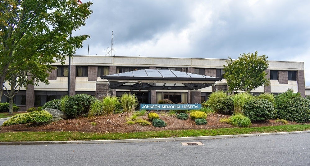
Diagnostic Imaging/Radiology at Johnson Memorial Hospital
Phone & Address
201 Chestnut Hill Rd
Stafford Springs
,
CT
06076
Get directions
860-684-8170
860-684-8499
Hide Map
Get directions