Interventional Pulmonology
Interventional pulmonology (IP), an advanced subspecialty of pulmonary medicine, focuses on using minimally invasive procedures and therapeutic techniques to diagnose and treat conditions in the lungs and chest. IP procedures yield exceptional outcomes and offer patients the potential advantage of avoiding more invasive surgery.
Our interventional pulmonologists, Anil Magge, MD and Amrita Karambelkar, MD, works hand-in-hand with an expert team of thoracic surgeons, oncologists, radiation oncologists and respiratory therapists to successfully manage complex airway and pleural diseases, including lung cancer and non-malignant diseases of the chest.
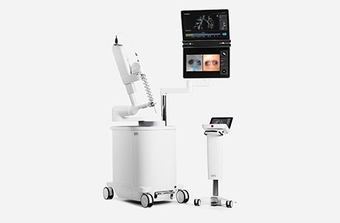
Ion Endoluminal System
Ion is used to perform robotic-assisted bronchoscopy. This system can reach all 18 segments of the lungs and be placed to obtain a biopsy. Ion is designed to address a challenging aspect of lung biopsy by enabling physicians to obtain tissue samples from deep within the lung.
Learn More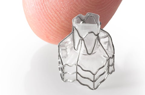
Endobronchial Valves
Endobronchial Valve Treatment is a minimally invasive treatment for people with severe COPD/emphysema. The valves are placed in selected airways during a bronchoscopy procedure (no incisions or cutting required) and are an alternative to the more invasive traditional lung volume reduction surgery.
Learn More

Saint Francis Hospital has become the first in Connecticut to combine Cios Spin, a state-of-the-art 3D imaging system, with the Ion endoluminal system for robotic bronchoscopy, enabling physicians to detect and diagnose lung cancer early.
Cios Spin, produced by Siemens Healthineers, is a mobile 3D imaging system, which allows doctors to pinpoint the exact location of a suspicious lung nodule. Prior to integrating the Cios Spin during an Ion procedure, a CT scan of the chest was taken to map a pathway to the nodule. By integrating Cios Spin with Ion, physicians are now able to navigate within the lung with even greater precision, with real-time, CT-like images.
During bronchoscopy with Ion, the physician uses a console to navigate to the target within the lung along a pre-planned pathway. The catheter can move 180° in any direction to pass through small, difficult to navigate airways to reach the lung nodule of concern. Ion’s peripheral vision probe provides direct vision during navigation. Once at the desired location, the catheter locks into place and biopsy tools are then passed through the catheter to take a sample of the nodule.
Specialized Care Programs
We offer state-of-the-art diagnosis and treatment and the highest quality of care for patients with advanced airway and pleural disease.
Central Airway Program
Obstruction and stenosis of the trachea and central bronchial airways can limit your ability to breathe and perform everyday tasks. This can happen with cancer where a tumor is compressing or growing within the trachea and bronchus and with non-cancerous obstructions such as stenosis or scarring. This can also be caused by infections, inflammatory diseases, or complications from treatments like endotracheal intubation and tracheostomies.
Our Central Airway Program removes obstructing tissue through bronchoscopic procedures like:
- Argon Plasma Coagulation
- Cryotherapy
- Cryospray
- Electrosurgery
- LASER
- Photodynamic therapy
- Rigid bronchoscopy
We can also place or remove airway stents (silicone, metallic, Y stents, iCast, Bioabsorbable, 3D customized stents), tracheostomy tubes, and Montgomery t-tubes/cannulas to help patients breathe comfortably.
Pleural Disease Program
The pleura is a thin sheet of tissue that surrounds the outside of your lungs. Pleural disease can cause fluid buildup in the chest, making it difficult to breathe which causes other symptoms like coughing and chest pain. This disease is a common condition for many patients with cardiac, liver, and renal disease, as well as numerous types of cancer.
In addition to fluid buildup, patients can have air leak into the chest cavity also known as pneumothorax.
Our program offers a comprehensive pleural care service and we perform in-clinic procedures to relieve patient symptoms and preventive measures to help keep patients out of the hospital. We perform:
- Thoracentesis
- Chest tube placement
- Tunneled or indwelling pleural catheters
For patients requiring further workup, patients can be taken to the operating room for:
- Medical thoracoscopy with pleural biopsies
- Pleurodesis
Tracheobronchomalacia Program
Tracheobronchomalacia is a condition where the cartilage in the trachea and bronchi are weakened, making it difficult to breathe. Tracheobronchomalacia symptoms include persistent cough, loss of breath, and recurrent respiratory infections.
Our Tracheobronchomalacia Program can help identify these conditions using state-of-art diagnostic procedures. We work closely with our interdisciplinary team to treat this condition with airway stents and collaborate with our thoracic surgery team to consider surgical approaches to stabilize the trachea and airways.

For more information about Trinity Health Of New England’s Interventional Pulmonology services, please call 860-714-4055 for Saint Francis Hospital or Saint Mary's Hospital and 413-748-9664 for Mercy Medical Center.