Vascular Medicine

Pioneer Valley Cardiology Associates is an IAC accredited facility for vascular diagnostic testing. Tests are performed by ARDMS registered vascular technologists and interpreted by by one of our cardiologists with training in vascular medicine. Physicians are on site at all times while tests are being performed. Our full spectrum of vascular diagnostic testing includes:
- Ankle Brachial Index (ABI):
Blood pressure in the arms is compared with measurements at the ankles and big toes. Blood pressure cuffs may also be placed at the thighs and just below the knees. This aids in diagnosis of peripheral arterial disease (PAD), a cause of leg pain. - Arterial Ultrasound:
Images of blood flowing through arteries are captured using sound waves. This is used to diagnose blockages in the legs, neck or kidneys. - Abdominal Ultrasound:
You will be asked not to eat anything on the day of this test to help us get good pictures. The technician will use sound waves to measure the size of your aorta. This is a screening test for abdominal aortic aneurysm, an enlargement of the main blood vessel running through the center of your body. - Carotid Intimal Medial Thickness (CIMT):
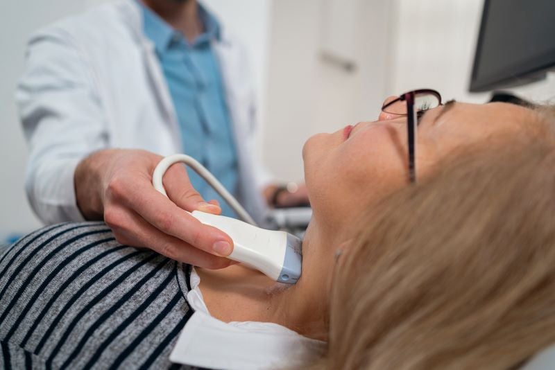
Your neck will be scanned with sound waves to measure the thickness of the inner lining of the carotid arteries. Your doctor may order this test to help determine the risk of developing cardiovascular disease. - Venous Ultrasound:
Leg veins will be scanned with sound waves. Deep vein thrombosis (DVT), or a blood clot in the leg, can be seen. Gentle compression of the veins allows us to see if they are functioning normally. This test also provides a roadmap of the veins in your legs, which is needed before having leg bypass surgery.
Based on the results of diagnostic testing, you may be referred to a vascular cardiologist or surgeon for consultation.
All procedures are minimally invasive with moderate sedation. There are no incisions and you don't need to be put completely asleep. Everything is done through a puncture in one of the arteries, typically in the leg. Through this tiny hole, catheters are placed into the artery and used to inject contrast dye. Live x-rays allow the doctor to see where the dye is going and to map your circulation. It is possible that you will have to spend a night in the hospital for observation afterwards.
- Angioplasty
Leg artery blockages are opened using high pressure balloons. These are rapidly inflated and deflated to crush plaque and open the blood vessel. This may be combined with drilling of plaque if the blockage is hard and made of calcium. Some balloons are coated with a special drug that gets implanted into the wall of the artery, keeping it from re-narrowing later on. - Stenting
Combined with angioplasty, a rigid metal scaffold (or stent) may be left behind to keep the artery open. Stents are permanent. They do not set off metal detectors and are safe for MRI. These are commonly used for arteries in the neck, kidneys and legs. - Aortic Endograft
An aortic aneurysm is a balloon-like enlargement of the biggest blood vessel in the human body. This can be treated with an endograft, which seals off the ballooned portion of aorta, stopping the blood pressure from reaching it and expanding it over time.


