Transesophageal Echocardiography (TEE)

Pioneer Valley Cardiology Associates has 4 cardiologists certified to perform Transesophageal Echocardiography (TEE) at Mercy Medical Center.
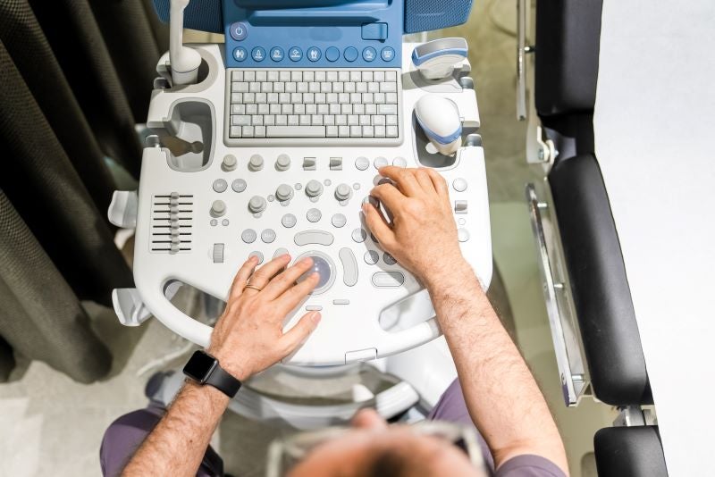
A transesophageal echocardiogram (TEE) is a test that produces detailed pictures of your heart's muscle, chambers and the arteries that lead to and from it using high-frequency sound waves (ultrasound). Different from a standard transthoracic echocardiogram during which images of the heart are obtained from moving a transducer externally to different locations across your chest and abdominal wall, a TEE is a specialized echocardiogram that obtains images internally. A transducer passes through the mouth, down the throat and into the esophagus, or food pipe. Very clear images of the heart and its structures can be obtained this way because the esophagus is so close to the upper chambers of the heart.
Doctors often use TEE when they need more detailed information than a standard echocardiogram is able to offer. For example, transesophageal echo is used to help surgeons prior to heart operations. We are able to provide 3 dimensional views with excellent anatomic detail, so the surgeon can plan his or her surgery prior to even cutting the chest! Furthermore, we can use TEE to help guide placement of valves during minimally invasive valve replacements. Also, there are structures that we cannot see well or at all from the chest wall. We use transesophageal echocardiogram when we need to see these structures. For example, we are often unable to see a structure called the left atrial appendage on standard echocardiogram. However, this structure is a common area for clots to develop. With the use of transesophageal echocardiogram we can assure no clots are present prior to putting the heart back into a normal sinus rhythm during a cardioversion procedure.
Other structures we may be able to visualize include:
- the thoracic aorta to look for dissections and aneurysms
- infections or 'vegetations' on valves, which can be life threatening
- congential anamolies-such as atrial and ventricular septal defects
The detailed images provided by TEE also help your cardiologist to diagnose:
- The size of your heart and thickness of its walls
- If there is abnormal tissue around your heart valves
- If blood is leaking backward through your heart valves (regurgitation) or
- If your valves are narrowed or blocked (stenosis)
- If blood clots are in the chambers of your heart
A transesophageal echocardiogram is performed in a procedure lab at the hospital. You should not drink or eat anything for several hours prior to the test. You will have an IV placed to administer fluids and medications. We will have you gargle several times with a numbing solution - typically a lidocaine solution. You will then be given some medications for sedation. For patients without many medical complications, this may be given by the cardiologist themselves. It could alternatively be performed by an anesthesiologist for patients with more involved medical issues, such as lung problems or chronic diseases. Either way, these medications will make you sleepy for the test. Once you are appropriately sedated, we will insert the transesophageal echocardiogram probe into your mouth, throat, then esophagus. Once it's in there, we mainly take pictures from the middle of the esophagus but will take pictures from the stomach as well. But well...a picture is worth a thousand words.
Generally, a transesophageal echocardiogram procedure is an outpatient one day procedure. It is low risk. Risk of trauma or serious injury to the esophagus is extremely rare. You may have a sore throat for a day or two after the procedure, which is normal. You will be monitored for 1-2 hours post procedure to assure you wake up and are able to take food and/or drink. You will then be able to go home, but will have to get a ride. You shouldn't be driving after receiving sedation medicine.